

Say Hi to Park Slope Eye’s new doc! Introducing Dr. Lisa DeClemente!

Laser scan

Transverse view of the retina with an OCT, image courtesy of Russell Neches
Occasionally I send patients to another clinic to get a laser scan of the back of their eye. What is this test, and why do we do it?
The official name of this procedure is called an “optical coherence tomography” scan or “OCT” for short. The machine emits a very short wavelength (~100 nm) laser beam that hits certain structures in the retina. The onboard processor is able to translate the information that the laser records into a topographical profile of whatever structure the doctor wants to see. This affords him the ability to notice any disruptions below the retinal surface.
I take retinal pictures of every single patient in order to examine key anatomical and functional components of the eye. Retinal photos are a brilliant form of technology that allows me to see very detailed things. Occasionally I also utilize special lenses to determine if a problem is present. However, these two techniques are sometimes not detailed enough to examine key hidden structures.
Instead of thinking about the retina, let’s use an apple analogy for a moment. If you take a picture of the side of an apple, you could see that it is red. You could see the outline and perhaps infer that it curves outward towards you. You wouldn’t be able to tell how deep the apple was or what the insides look like. Taking an OCT of the apple would be like cutting it in half and observing a cross section. You would now be able to see that red color is actually a very thin skin. You could also measure the depth of the apple, see that the majority of it’s volume is composed of a fibrous network, and notice a small cavity in the center that’s filled with seeds. If the seeds were displaced to the side, a traditional camera picture wouldn’t notice anything out of the ordinary since the external appearance is untouched. However, an OCT would be able to tell that they are not in their proper position. For almost every patient, a retinal photo is way above the standard of care that’s required to examine their eyes. However, some patients’ eyes either present with abnormalities or have a suspicious appearance. Those patients require the detailed level of imaging and analysis that an OCT can provide.
It’s important to clarify a few misconceptions about getting an OCT:
-Does it hurt?
It doesn’t hurt at all. It’s actually slightly more gentle than a camera flash
-Will it fix my problem.
No. There are many types of lasers that doctors use on the eye. An OCT does diagnostic imaging only. That means that it shows the doctor what is the issue and does not fix it.
-Why didn’t my doctor see what the laser saw?
A doctor only orders an OCT when a known or suspected problem lies in an area of the retina that’s not visible to human eyes and traditional machines. Unless your doctor has x-ray vision, he won’t be able to see the deep hidden retinal layers.
-Come on doc. Is this really important?
Absolutely! With many retinal pathologies the OCT reading is how the doctor determines what treatment or monitoring protocols he will use. Additionally, he now has a snapshot of the retina at a given time and can track it’s evolution in the future.
-Why doesn’t my doctor have an OCT?
Most patients don’t require such a scan. Typically I will see only 1 patient who needs an OCT each week. Although I can perform the scan and interpret the results with ease, I prefer my patients to be scanned by a doctor who does 10 a day and specializes in that type of care.
-You’re sending me to a disease specialist? I must have something really bad.
Needing an OCT does not mean that you have a serious disease. It does mean that your eyes present slightly atypically and an extra scan is the prudent thing to do. Frequently the doctor who does my scanning will say, “It looks okay. We’ll just monitor it. Go see Dr. Ritenour in a year.” Some patients I know are healthy, but have an elevated risk for developing a problem in the future. In theses cases, the OCT scan is a baseline “snapshot” that we can always compare back to in the future.
Leave a comment or email me if you have any questions or concerns.
Spencer Ritenour, O.D.
dr.ritenour@parkslopeeye.com
What is 20/20?
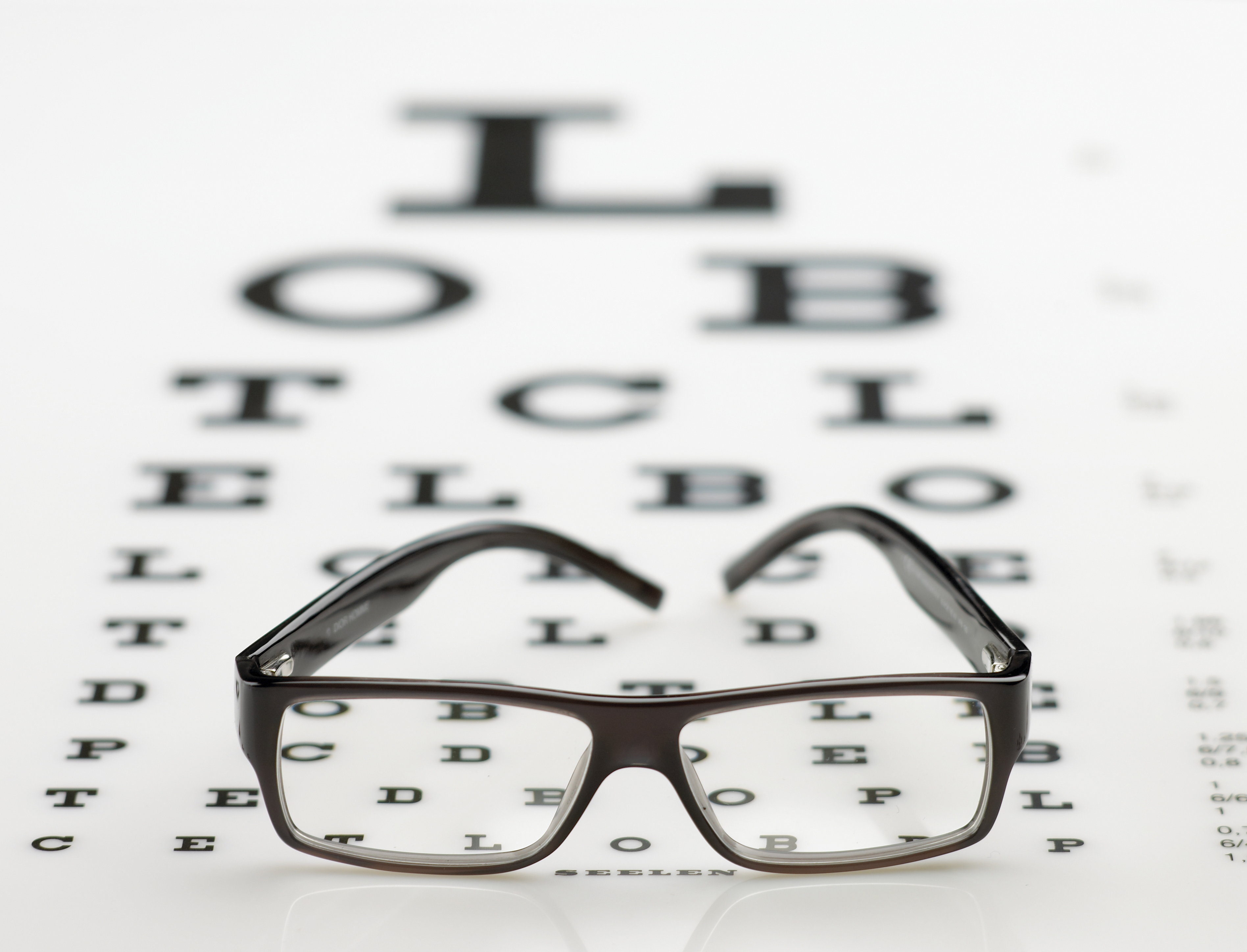 Optometrists love to toss around the term 20/20: “You have 20/20 vison.” or “Gosh, you’re seeing better than 20/20.” Perhaps, you use it at home: “How does this dress look?” “Sorry honey – I don’t see 20/20.” The news program “20/20” was actually named after the visual measurement. Everyone knows 20/20 is ideal, but few actually know what it means.
To know how well someone sees, we must use a standardized system. This affords doctors the ability to know if someone needs more or less correction and to compare the vision between members of a population. For example, it’s means very little to accurately check the vision of an eight month old girl and then record it as “pretty good” or “seems normal.” It’s far superior to be able to record her vision as 20/60 or 20/80. Then visual changes may be tracked with tighter accuracy, her visual acuity may be compared against what’s normal for 8 month olds, and her vision may be corrected or observed against a quantifiable system.
The 20/xx system is also known as the Snellen visual acuity system. The top number refers to the testing distance in feet. Twenty feet is considered to be “optical infinity” due to how our eyes focus and the behavior of light waves at this distance. However, most offices do not have patients sit 20 feet from a chart due to space constraints. Through the use of mirrors and chart calibrations, the shorter distance is compensated for remarkably well.
The bottom number is a bit more complex. It’s the distance at which each element of a letter has an angular height of 1 minute of arc (or 1/60th of a degree). The letter “E” on an eye chart has 5 elements. There is the top bar, a space, the middle bar, another space, and the bottom bar. When one stands 400 feet from the big E at the top of the chart, each of these elements subtends 1 minute of arc. Therefore the size of the letter is called 20/400. For a letter on the 20/20 line, one must stand 20 feet away for each element to subtend 1 minute of arc.
The neurological processing in the human eye and key areas of the brain allows people to easily discriminate between letters with 1 minute of arc (or smaller) features. With a (compensated or actual) testing distance of 20 feet, optometrists therefore embrace the 20/20 line as a perfect endpoint. Please don’t fret if you can’t see 20/20 without glasses or contacts. That will be the subject of a blog post in the near future. Seeing 20/20 with or without visual correction means that at 20 feet away, your eyes see as well as they are expected see. If you can’t read 20/20, please schedule an appointment, and I’d love to help boost your vision.
Spencer Ritenour, O.D.
dr.ritenour@parkslopeeye.com
…but my vision isn’t blurry.

How often do you visit your eye doctor? Annually? Every 5 years? Still haven’t been checked? Perhaps a better question is how often should you visit your eye doctor. For the general population, optometrists recommend a visit at least every 12 months. Still it’s common for people to wait until their vision gets blurry. On the surface that seems sensible, but it overlooks some very important elements of good eye health.
Every eye exam does include a determination of one’s glasses and/or contact lens prescription. While this is important for good vision, it’s just a small component of the examination. When light enters the eye, it gets refocused no less than 12 times before being converted to an electrical signal behind the eye. In most people, the summation of these focusing steps is not quite perfect – leading to a prescription that compensates for the difference. This would be the end of the story if the eyes were simply glass and electronics like a camera lens. Obviously they are living tissue that have special needs and interacts with the rest of the body.
 There are disorders that can affect all of the elements that comprise the eye (e.g. the eyelid, tears, lens, retina). Eye specific diseases include glaucoma, cataracts, macular degeneration, and many more. Many systemic conditions such diabetes, hypertension, and high cholesterol may cause ocular complications as well. In fact in certain instances, an optometrist is the first doctor to see these signs in a patient. During a eye doctor’s examination he observes the external eye structures under high magnification. Also he will look inside the eye at the all internal structures. Any abnormalities are treated or aggressively monitored as warranted. In addition to examining the physical structure of the eye, the physiology or functioning of the eye is equally important to check. He will ensure that they eyes are moving properly, working well together and independently, communicating with the brain, and being supplied with blood as they should.
Some people are fortunate enough to not need glasses until about age 40 or maybe never need them. Other people notice that their prescription stops changing in their late teens or early twenties. Regardless of how blurry or clear a person’s vision is, it’s very important to ensure that her eyes are structurally sound and functioning to the max. If a problem is ever detected, it is far superior to catch it in the early stages than to catch it much later. Don’t let “good enough” vision rob you of a lifetime of stellar vision and healthy eyes.
Spencer Ritenour, O.D.
dr.ritenour@parkslopeeye.com
Photo sesh.

Funduscopic photo of a patient's retina
Bored with your bar pics? Had it with holiday photos?
At Park Slope Eye we take pictures of the inside of the eye with every full exam. Our clinic uses a professional grade Canon DSLR with a special ophthalmic attachment that allows us to take amazing high quality shots. Also, our exam room is equipped with a high res monitor where we display every photo. Dr. Bazan and I show every patient exactly what their retinas look like and thoroughly explain how they’re functioning. If any pathology is discovered, the pictures assist us in aggressively monitoring and treating the condition. We both strongly believe in making sure that our patients are highly educated about their health. The pictures allow you to see exactly what we can see. We can even email them to you if you like. They make great Facebook profile pics!
Eric I. (5/5) on Yelp
quoted from Eric I. (5/5) on Yelp
Screw hanging at coffee shops to write the next great american novel. Park Slope eye has free coffee (you can even take the mug home) , snacks and they have a computer for your use so you could write…
Thanks for also checking in! You are the man!

I was recently listed on this site. I really think they have the right mindset and wish them great success. WellnessCenters.com’s mission is to empower millions of individuals to find their perfect well-being experiences! Try them out.
![]()
Contact Lens Saftey
Contact Lens Safety
If you aren’t able to throw out your contacts after each use (daily disposables!) please take heed to what the FDA has to say here: http://www.fda.gov/ForConsumers/ConsumerUpdates/ucm164197.htm
Park Slope Eye is located in Brooklyn, NY. For more info contact Justin Bazan, OD, the optometrist of Park Slope Eye, at Dr.Bazan@ParkSlopeEye.com or visit www.ParkSlopeEye.com Also, check us out on Yelp!, Twitter and FaceBook.
![]()

